Original Article
, Volume: 11( 2)Bacterial Synthesis and Applications of Nanoparticles
Ayesha A*
Institute of Molecular Biology & Biotechnology, The University of Lahore, Pakistan
- *Correspondence:
- Ayesha A, Institute of Molecular Biology & Biotechnology, The University of Lahore, Punjab Pakistan, Tel: 0322-4656026; E-mail: Ashu.arshad113@gmail.com
Received: Aug 14, 2017; Accepted: Sep 09, 2017; Published: Sep 16, 2017
Citation: Ayesha A. Bacterial Synthesis and Applications of Nanoparticles. Nano Sci Nano Technol. 2017;11(2):119
Abstract
Nanoparticles synthesis is the real division in the area of relevant Nanotechnology and Nanoscience. As of late, the merging amongst nanotechnology and science has made the new field of Nanobiotechnology that joins the utilization of natural elements, for example, algae, microscopic organisms, parasites, infections, yeasts and plants in various biophysical and biochemical procedures. The natural combination forms have a critical prospective to support nanoparticles generation without the utilization of brutal, harmful and costly chemicals usually utilized as a part of ordinary physical and substance forms. Combination of nanoparticles (NPs) utilizing microscopic organisms has risen as quickly creating research range in nanotechnology over the globe. The procedures of NPs combination result with required shapes and controlled sizes, quick and clean. These days, a variety of nanoparticles with very much characterized synthetic organization, size and morphology have been combined by utilizing distinctive microorganisms and their applications in numerous mechanical fields have been investigated. The uses of these biosynthesized nanoparticles in a wide range of potential zones are exhibited including focused on targeted drug passage, malignancy treatment and DNA investigation, biosensors and magnetic resonance imaging (MRI). The consumption of microorganisms for nanoparticles synthesis is a genuinely unique range of examination with extensive prospective for more improvement.
Keywords
Nanoscience; Nanotechnology; Nanomaterials; Biological synthesis; Bacterial synthesis
Introduction
Nanoparticles, has gotten specific interest for a wide variety of fields. Nanoparticles are characterized as particulate scatterings of strong particles with no less than one measurement of 10 nm to 1000 nm in size. The mainly vital element of NPs is the surface region to volume perspective proportion, permitting to cooperate with different particle simpler.
Nanoscience and nanotechnology has pulled in an extraordinary interest in the course of the most recent couple of years because of its potential effect on numerous logical ranges, for example, strength, direction, pharmaceutical commercial enterprises, hardware and space businesses. This innovation manages little structures and little estimated materials of measurements in the scope of couple of nanometers to fewer than 100 nanometers. NPs show exceptional and extensively changed substance, physical and organic properties contrasted with main part of the same synthetic organization, in view of their high surface-to-volume extent. These particles moreover have various applications in different fields, for instance, healing imaging, nanocomposites, channels, drug transport and hyperthermia of tumors. A key domain of examination in nanoscience discussed the combination of nanometer range particles of diverse shapes, monodispersity and sizes [1-33].
Experimental
Morphology of nanoparticles
The morphological uniqueness of nanoparticles is: flatness and aspect ratio.
1- High aspect ratio NPs.
2- Small aspect ratio NPs.
Nanomaterials types
NMs might be natural, inorganic and composite in nature. Natural nanomaterials incorporate man-sized proteins, nucleic acids and carbon nanotubes and so forth. On the other side inorganic nanomaterials incorporate metallic NPs, mixes and mass nano-organized materials.
These metallic nanomaterials are Ag, Au, Pd and Pt [1,2]. It likewise incorporates bimetallic nanoparticles like Ag-Pt, Ag-Au, Pd-Au, Ti [TiO2], oxides of Fe [Fe2O3-] magnetite, Fe3O4-magnetite [3], Zn [ZnO, Si [SiO2] and in addition sulfides of various metals [CdS [4], FeS [3], ZnS [5,6]. Different nanomaterials incorporate mass nano-organized metals, precious stones and powders of Te [7], Ti [8], Se, Al [9] and move metals like Ni, Co, Cu, Cr, Zr [10] and Pb. Then again, nano-composite materials include quantum Dots, carbon nanotubes [11], nano-shells, nano-bars, nano-wires, nano-gels and nano-emulsions and so on. Nanoparticles can be sorted by measurements for instance zero-dimensional iota groups, one-dimensional regulated multilayered, two-dimensional NPs, three-dimensional, ultra-fine grained over layers, [12].
NMs properties
The nanoscale is unique in light of the fact that no strong can be readied littler than it. It is likewise remarkable in light of the fact that a large portion of the components of the physico-synthetic and natural world capacity on estimation scales going from 0.1 nm to 100 nm. By and large, the lessening in size of the particles builds their surface to volume proportion which in this way expands their reactivity.
At nano-scale, particles of various components show amazing physical, compound and natural properties. In contrast with traditional coarse-grained partners, NPs show lower softening point, electrical resistivity, particular warmth, diffusivity, pliancy and mechanical quality alongside changes in their electromagnetic and synergist properties. Researchers are attempting to decipher these properties connected with NPs in different advancements.
Synthesis of nanomaterials
The nanomaterials synthesis involves chemical, physical and biological methods. Biological procedures are still in the stage of development [13] Figure 1.

Figure 1: Different methods for the synthesis of NPs.
Physical and chemical synthesis of nanomaterials
Physical methods for blending nanomaterials include crushing, physical vapor deposition (PVD), ball processing, lithography and pyrolysis. Among these, pounding and pyrolysis have been utilized most regularly. While, there should arise an occurrence of substance systems including electrolysis, sputtering, sol gel synthesis, CVD (chemical vapor deposition), sputtering and inactive gas buildup have been more mainstream. These types of techniques are advantageous however they are dangerous as well furthermore exorbitant.
Biological synthesis of nanomaterials
Biological sources include plants and microbes like yeast, fungi, bacteria and actinomycetes. It has been found that biological sources act as reducing metal ions from the soil. This process is quite safe as comparison to other physical and chemical methods. It is a lesser amount of energy concentrated loom but the nanomaterials produced by these methods have found very reactive. This “enzymatic” proposal is extra workable by the reality that most of the microorganisms raise at climate situation of temperature and pH (Table 1).
| Technique | Advantages | Disadvantages | Chemicals/ Microorganims used |
References |
|---|---|---|---|---|
| Physical | Firm join,baffled indata andeffectiveness note being of NPs Deliver of the crystal expansion, synthesis of the particles |
Mandal et al.[9] | ||
| Chemical | High monodispersity (5–15%) |
Costly feeble-minded in figure and sortie consequently claim of mortal chemicals Note environmental easy to deal with resolution of NP Manage of the crystal development, synthesis of the particles Low yield |
Reducing agents, such as methoxypolyethylene glycol, sodium borohydride, potassiumbitartarate, hydrazine Stabilizing agents, such as polyvinyl pyrrolidone, sodium dodecylbenzylsulfate. |
Sastry et al. Mandal et al. [9] |
| Biological | Low monodispersity (~40–50%), environmental friendly |
Yeast, bacteria, fungi, algae, plants, viruses and actinomycetes. |
Sastry et al. |
Table 1: Advantaged and disadvantages of different techniques.
| Nanoparticle | Applications | Toxicity | References |
|---|---|---|---|
| Titanium dioxide | Titanium nanoparticles have been applied in the pharmaceutical industry as drug delivery vehicles and in excipient formulations. | The composite material combines the high adsorption capability of apatite with the photocatalytic activity of titanium. Apatite coatings may thus become useful in the attenuation of the toxicological effects of inorganic metal oxide nanoparticles. | (Acosta-Torres et al. [62]; Driscoll et al., [63]Salata et al. [64]Cantado et al. 2008;[65]Guo et al. [66]; Sikong et al.[67] |
| Iron oxide | Used in cellular therapy, such as cell labeling and targeting and as a tool for cell biology research to separate and purify cell populations. Also used in: tissue repair; drug delivery; magnetic resonance imaging; hyperthermia; magnetofection. |
No toxicity reported. | (Acosta-Torres et al. [62] Arbab et al. [68]Sanpo et al. [19] |
| Silver | Used for covering urinary catheters, surgical instruments and bone prostheses. Additionally, silver has been used in water and air filtration to eliminate microorganisms. AgNPs have been added to soft tissue Conditioners for prosthetic devices |
Exposure of metal-containing nanoparticles to human lung epithelial cells generates ROS, which can lead to oxidative stress and cellular damage (AgNPs). Silver nanowires resulted in the strongest cytotoxicity and immunological responses, whereas spherical silver particles had negligible effects on cells when tested in human cells. |
(Limbach et al. [69] Niño-Martínez et al. [70] Ahn et al. [71] |
Table 7: Nanoparticle applications and their toxicity.
Synthesis of nanoparticles can occur either intracellularly or extracellularly. Intracellular synthesis of nanoparticles requires extra steps, for example, ultrasound treatment or responses with appropriate cleansers to discharge the combined nanoparticles [14-16].
In the meantime, extracellular biosynthesis is inferior and it requires more straightforward downstream handling. As a result of this, numerous studies were focused on extracellular techniques for the blend of nanoparticles (Table 2).
| Biological Entity | Microorganisms | Types of NPs synthesized | References |
|---|---|---|---|
| Bacteria 1 nm–200 nm diameter | Bacillus licheniformis;Bacillussubtilis; Bacillus stearothermophilus; Clostridium thermoaceticum; Desulfobacteriaceae; E. coli; Klebsiella aerogenes; Klebsiella pneumonia; Lactobacillus increase; Rhodopseudomonas capsulata; Magnetospirillummagnetotacticum; Desulfovibrio desulfuricans; Pseudomonas aeruginosa; Pseudomonas stutzeriAG259; Rhodopseudomonas palustris; Thermoanaerobacter ethanolicus (TOR-39) | Ag, Au, CdS, pd, Fe3O4, ZnS | [4,9,15,16] |
Table 2: Microorganisms involve in the synthesis of different NPs.
Why choose bacteria?
Nanoparticles are constantly being studied and developed. Three methods are used to synthesize the nanomaterials. Physical methods include lithography, pyrolysis, vapour pressure etc. but these methods are quite expensive. It is important that physical and chemical methods are low yield, energy intensive, difficult to scale up, often produce high levels of hazardous wastes and may require the use of costly precursors.
Besides, a chemical method includes Irradiation reduction, micro-emulsion method and electrochemical reduction but chemical methods are quite hazardous this synthesis may still lead to the existence of some lethal chemical species adsorbed on the surface that may have unpleasant effects in medical applications.
Hence, there is a need to develop inexpensive, clean, nonlethal and environmentally kind synthesis methods. Therefore, in last year’s researchers have turned to biological systems for inspiration in nanoparticle synthesis. Microorganisms are recently found as possible environment friendly nanofactories, even though they have many biotechnological uses such as remediation of lethal metals.
It has confirmed that prokaryotes earnest powerfully as action of synthesizing nanoparticles. Suited to their over-sufficiency in atmosphere and their gift to accommodate to original broadcasting situation, bacteria are a good selection for research. Bacteria are in addition remaining maturation, economical to grow and easy to control. Heaping up diffusion such as oxygenation, incubation time and temperature can be easily manipulated. It was discovered that irregular the pH of the collecting operation by means of incubation mean in the play of nanoparticles of incompatible display and suit, predominant such transfer is standard, as unconditional morphologies of NPs are necessary for different applications such as catalysts, optics or anti-microbials.
NPs synthesis by bacteria
Bacteria have the remarkable ability to reduce and synthesize heavy metals and nanomaterials. Some bacterial species have developed the ability to remedy to specific defense mechanisms to control stresses like toxicity of nanomaterials. Research has paying attention heavily on prokaryotes as a means to synthesize nanoparticles (Table 3).
| Bacteria | Nanoparticle | Size | Morphology | Reference |
|---|---|---|---|---|
| Aeromonas sp. SH10 | Silver | 6.4 | Rai et al. [17] | |
| Bacillus Cereus | Silver | 20–40 | Spherical | Sunkar et al. [18] |
| Bacillus megatheriumD01 | Gold | 1.9 ± 0.8 | Spherical | Wen et al. [19] |
| Bacillus subtilis | Silver | 5–50 | Triangular and spherical | Beveridge et al. [20] |
| Clostridium thermoaceticum | Cadmium sulfide | Amorphous | ||
| Desulfobacteraceae | Zinc sulfide | 2–5 | Spherical | |
| Desulfovibrio (desulfuricans,vulgaris, magneticus strain RS-1) |
Palladium, selenium, gold, uranium, chromium and magnetite | Up to 30 | Crystalline | |
| Escherichia coli (DH5?, MC4100) | Silver, gold | Less than 10-50 | Spherical, triangular, hexagonal and rod shape |
Mahanty et al. [21] |
| Geobacillussp. | gold | 5-50 | Quasi-hexagonal | |
| Klebsiella (aerogenes, pneumonia) | Cadmium sulfide, silver | (average size of 52.2) | Spherical | Shahverdi et al.[22] |
| Lactobacillus strains | Gold, silver, gold-silver alloys, titanium | 100-300 | Crystalline and cluster | |
| Magnetospirillum Magnetotacticum |
Magnetite | Cluster | ||
| Nocardiopsissp. MBRC-1 | Silver | 45 | spherical | Manivasagan et al. [23] |
| Pseudomonas (aeruginosa, fluorescens, putidaNCIM 2650, stutzeriAG259) | Gold, silver, lanthanum | 35–46 and up to 200 |
Crystalline silver, Hexagonal, equilateral triangle, and monoclinic silver |
|
| Rhodobactersphaeroides | ZnS | Average diameter of 8 |
Spherical | |
| Rhodopseudomonas (capsulate, palustris) | Gold, CdS | 8.01 ± 0.25 | Crystalline, FCC | Bai et al. |
| Shewanella algae (strain BRY, putrefaciens (Gs-15) ) |
Gold, magnetite | Various morphologies altered with pH |
Konishi et al. [15] | |
| Thermoanaerobacter ethanolicus TOR-39 |
Cobalt, chromium, magnetite and nickel | octahedral | Rai et al. [17] |
Table 3: Bacteria involve in synthesis of different nanoparticle.
Due to their plenty in the surroundings and their ability to adjust to extreme conditions, bacteria are a good option to learn. They are also fast growing, cheap to develop and easy to control. Growth conditions such as temperature, oxygenation and incubation time can be easily controlled.
Synthesis Types
Synthesis is of two types; intracellular and extracellular.
Intracellular nanosynthesis
A large number of bacterial species have been investigated for intracellular bio-nanosynthesis of metallic and non-metallic nanoparticles the details are as under;
Intracellular synthesis of metallic nanomaterials
Intracellular metal nanoparticles combine has been accounted for in different microorganisms as bio-nanofactories [25,26]. Generally, both bacterial synthesis indicated has been very important part in bio-geo cycles. This capacity has helped them in change of different mixes and basic metal in nature. Profoundly lethal centralization of metal particles is detoxified by microscopic organisms through lessening or oxidation, precipitation or complexation, transportation or efflux frameworks and so on. These properties have marked them as potential bioremediation specialists in soil and oceanic situations. In this point of view, microscopic organisms part in biogenesis of nanoparticles has additionally been explained last couple of decades. In this manner, the synthesis of intracellular metal nanoparticles including gold, silver, gold-silver alloy, platinum, palladium, uranium has been researched in a few bacterial strains.
Gold nanoparticles:Bacillus subtilis 168 encouraged gold (Au+3) particles to Au0 nanoparticles with octahedral morphology has been accounted for onto their cell dividers [26]. Another bacterium, Geobacterferrireducens diminished Au particles in periplasmic space to deliver AuNPs. Shewanella algae likewise decreased Au+3 particles to natural AuNPs at 25°C in periplasmic space and on its cell surface. Under various pH conditions distinctive sizes of NPs were watched [15].
A cyanobacterium, Plectonemaboryanum UTEX485, synthesized AuNPs at 25 to 200°C with the assistance of some external layer proteins, lipopolysaccharides and phospholipids. This blend has been connected with indicated detoxification systems in microscopic organisms [25]. Morphologically, round, triangular or hexagonal AuNPs has been bio-diminishment of wrote about cell surface in E. coli DH5α with morphologies. Besides, this synthesis has been connected with plasma layer related NADPH-subordinate proteins and carotenoids in Rhodobactercapsulatus intervened biosynthesis of AuNPs.
Silver nanoparticles:Comprehensively, AgNPs synthesis has been connected with bacterial cell surfaces. If there should be an occurrence of Pseudomonas stutzeri AG259 crystalline of silver sulfide acanthite (Ag2S) were situated in periplasmic space with monoclinic, triangular, equilateral and hexagonal morphologies. It was connected with cell surfaces which were at first connected with biosorption and afterward diminishment Ag+ particles to frame AgNPs in Lactobacillus sp. A09 at 30°C [26]. For the arrangement of AgNPs, the nucleationdestinations are thought to be given by silver-restricting proteins which give amino corrosive moieties. Silver precious stones with face-focused cubic morphology were accelerated by AG3 and AG4 (silver encouraging peptides). Bacillus sp. additionally integrated AgNPs in its periplasmic space. This aggregation or precipitation of Ag at cell divider at temperature 60°C has likewise been considered as a detoxification procedure encouraged through periplasmic proteins [5].
Other metals:Synthesis of bimetallic NPs has likewise been accounted for in couple of microscopic organisms, Biogenesis of silver, gold and gold-silver composites was examined by utilizing B. subtilis[20], P. stutzeri, Lactobacillus sp,Corynebacterium sp. [5], sulfate-diminishing microscopic organisms [25], P.boryanum [26], E.coli, R.capsulatus and Bacillus sp. with circular, hexagonal and cubic morphologies. Intracellular bacterial synthesis of platinum (Pt) NPs was seen inS. algae in its periplasmic space amongst inward and external films. Palladium (Pd) NPs were intracellularlysynthesised by Desulfoviriodesulfuricans NCIMB 8307which diminished Pdnanocrystals on its cell surface with the assistance of formate (electron contributor). In a comparative study, S. oneidensis MR-1 was uncovered to frame Pdnanocrystals inside periplasmic space and in addition on the cell divider [27]. Uranium nanocrystals as urinate (UO2) were observed to be encouraged by Desulfosporosinus sp., on its cell surface, which can be useful in minimizing the defilement of solvent radionuclides in soils and dregs by changing the dissolvable to insoluble structure.
Magnetic nanomaterials:Intracellular bacterial biosynthesis of attractive nanocrystals of magnetite (Fe3O4) nanoparticles was observed to be shown by different microscopic organisms including Aquaspirillummagnetotacticum[3], Magnetospirullum magnetotacticum, M. magnetotacticumMS-1, M.gryphiswaldense, CandidatusMagnetoglobusmulticellularis, Magnetotactic bacterium MV-1[28], Sulfatedecreasing microscopic organisms. Biomineralized magnetite NPs are generally synthesised by magnetotactic microorganisms which have particular structures called magnetosomes in their phones. These magnetotactic microscopic organisms live in marine and new water silt and deliver intracellular, layer bound magnetite, pyrrhotite, ferromagnetic iron sulfide, greigite (Fe3S4) [28] and at some point non-attractive mineral (e.g. iron pyrite) as chains [5]. The attractive NPs are isolated from arrangement by utilizing high-angle attractive separators. The magnetosomes create both crystalline and non-crystalline attractive nanocrystals of prevalent morphologies including octahedral, exceedingly requested single or different chains, unpredictable faceted cub-octahedral shape, parallelepiped, octahedral or hexagonal crystal-like shapes, tied down and collected in phospholipids layers of microbes.
Sulphidenanomaterials:The semiconductor nanocrystals of cadmium sulfide (CdS) were intracellularly incorporated by different microorganisms including, Klebsiella pneumonia[29],Clostridium thermoaceticum and E. coli[4]. It was seen that, CdS nanocrystals were accelerated by the activity of cysteine desulfhydrase which brought about desulfhydration of cysteine either on cell surfaces or in medium. These bio-interceded semiconductor CdS nanoparticles displayed optical and photoactive properties and indicated round and circular shapes. In another study, round NPs of ZnS were naturally orchestrated by organisms having a place with Desulfobacteriaceae.
Non-metallic nanomaterials
Bionanosynthesis of non-metals like selenium have likewise been examined. Different microscopic organisms including Stenotrophomonasmaltophilia SELTE02, Enterobactercloacea SLD1a-1, Rhodospirullumrubrum, Desulfovibriodesulfuricans, E. coli [30], P. stutzeri, Tetrathiobacterkashmirensis, P. aeruginosa SNT1 have been found to store selenium NPs after bio-decreasing selenite to essential selenium in cell cytoplasm, periplasmic space andextracellularly with various shapes like granular, circular, fibrillar or in totals [31] (Table 4).
| Organism | Metal/Non-metal | Size (nm) |
Location of synthesis | Shapes | References |
|---|---|---|---|---|---|
| Idiomarinaspp. PR58-8 | Ag | 26 | Intracellular | ||
| Pseudomonas spp. | Ag | 156-265 | Intracellular | Thomas et al. [30] | |
| Bacillus subtilis 168 | Au | 5–25 | Intracellular | Octahedral | Beveridge and Murray [20] |
| Shewanella algae | Au | 10-20 | Intracellular | Lengkeet al.[25] | |
| Plectonema boryanumUTEX485 |
Au | 10 | Intracellular | Cubic | Lengkeet al. [25] |
| Escherichia coli DH5a | Au | Intracellular | Spherical | ||
| P. stutzeriAG259 | Ag, Ag2S | 200 | Intracellular | ||
| Corynebacterium spp. SH09 |
Ag | 10-15 | Intracellular | Zhang et al. [5] | |
| Bacillus spp. | Ag | 5-15 | Intracellular | ||
| Lactobacillus spp. | Au, Ag, Au–Ag | 20-50 | Intracellular | Hexagonal | |
| P. aeruginosa SNT1 | Se | Intracellular | Spherical | Yadav et al. [29] | |
| Desulfovibrio desulfuricans |
Pd | 50 | Intracellular | ||
| S. oneidensisMR–1 | Pd | Intracellular | De Windtet al.[27] | ||
| Aquaspirillum Magnetotacticum |
Fe3O4 | 40-50 | Intracellular | Octahedral | Mann et al.[3] |
| Magnetotactic bacterium MV-1 | Fe3O4 | 40×40×60 | Intracellular | Parallelepiped | Bazylinskiet al.[26] |
| M.gryphiswaldense | Magnetite | 35-120 | Intracellular | Cubo-octahedral hexagonal |
Table 4: Intracellular synthesis of nanoparticles by bacteria.
Extracellular nanosynthesis
A number of bacteria have been studied for their potential of extracellular bionanosynthesis.
Extracellular synthesis of metallic nanomaterials
Gold nanoparticles: Bioreduction of Au+3 into metallic AuNPs was seen by Rhodopseudomonascapsulata at room temperature. The particles were obtained with round and triangular morphology. Arrangement of various sizes and states of these NPs were discovered pH division of the response mixture.
Besides, nearness of diminishing and topping proteins of 14kDato 98 kDa size was confirmed through SDS-PAGE investigation. Iron (Fe+3) thinning archea and bacteria, for example, Pyrobalaculumislandicum, S. algae, G. sulfurreducens,Thermotogamaritimand Pyrococcusenraged, interceded precipitation of gold from ionic to metallic structure within thesight of hydrogen. Au+3 reductases close to the cell surfaces brought on this precipitation. P. aeruginosa (ATCC 90271, strain1 and strain 2) interceded synthesis of AuNPs was uncovered. Because of surface plasmon sound of greater NPs, medium shading changed from pink to blue.
B. megatherium D01 in dried structure synthesized spherical AuNPs at 26°C by reducing gold salts by utilizing dodecanethiol as topping specialist for balancing out NPs furthermore adjusting size, shape and monodispersity of NPs [20]. The morphologies of AuNPs manufactured by P. aeruginosa, R. capsulataand B. megatherium D01 were observed to be nanowires triangular and round fit as a fiddle extending in size from 1.9nmto 400 nm.
Silver nanoparticles:Dried cells of Aeromonas sp. SH10 were utilized to deliver monodispersed AgNPswith uniform size and security. A fast combination of AgNPs was seen by bioreduction of silver (Ag+) particles to metallic Ag0 by the activity of society supernatants of E. coli, Enterobacter cloacae and K. pneumonia[23]. The bioreduction was hindered when piperitone was added to the response blend which affirmed the nearness of nitro-reductase. Extracellular amalgamation of AgNPs was additionally seen by B. licheniformis[33].
Biogenesis of AgNPs was additionally exhibited in cellulose layers of Acetobacterxylinum when bacterial societies were presented to the arrangement containing Ag+ particles and tri-ethanol-amine (Ag+-TAE). Extracellular blend of AgNPs with round morphology was seen by microbes, for example, E. cloacae, Aeromonassp. SH10, E. coli, A. xylinum, B.licheniformis, Morganella sp. and K. pneumonia.
Other nanoparticles: Nanoparticles of platinum were accounted for extracellularly by the activity of different microscopic organisms, for example, P. boryanum UTEX 485 and Cyanobacterium with different morphologies containing dendritic, circular and dab like chains [25]. P. boryanum UTEX 485 integrated PtNPs at 25 to 100°C. Round molded NPs of titanium as totals were combined extracellularly by Lactobacillus sp. society filtrate [8].
Acellular concentrates of Micrococcus lactilyticus accelerated uranium NPs by lessening dissolvable U6+ to insoluble U4+[34]. Alteromonasputrefaciens created uranium NPs responded with hydrogen (electron giver) and U6+ (electron acceptor).
Uranium NPs were delivered by G.metallireducens GS-15 when under anaerobic conditions acetic acid derivation (electron benefactor) and U6+ (electron acceptor) did bioreduction of uranium particles. Because of nearness of MtrC (c-sort cytochrome) on outside layer of S. oneidensis MR-1, it did bioreduction of uranium particles with UO2-EPS (polymeric substance) extracellularly and in addition in periplasm.
Magnetic nanomaterials: Extracellular biosynthesis of magnetite NPs has been done by some non-magnetotactic microorganisms. Geobactermetallireducens GS-15, a non-magnetotactic bacterium disconnected from waterway base, created ultrafine magnetite NPs by lessening ferric oxide. In this procedure ferric particles (electron acceptors) responded with natural matter (electron benefactors).
Magnetite nanoparticles with octahedral morphology were incorporated extracellularly at 25°C by another bacterial strain named as TOR-39. Microscopic organisms were noted to go about as biocatalysts for precipitation of magnetite NPs. Ferric reduction microscopic organisms, TOR-39 andThermoanaerobacterethanolicus intervened electrochemical response which octahedral move metals (Ni, Cr, Co) substituted attractive nanocrystals were integrated.
The creation of semi round magnetite nanocrystals were exhibited by [8] by utilizing Actinobacter sp. (non-magnetotactic bacterium). Magnetite NPs with semi round and octahedral morphologies were incorporated extracellularly by TOR-39, G.metallireducensandActinobacter sp. [8]. Cubic spinel-molded single-crystalline ferromagnetic Co3O4 nanoparticles were synthesised by Brevibacteriumcasei[35] which is a metal-tolerant bacterium by utilizing watery cobalt acetic acid derivation as antecedent.
Sulfide nanomaterials: Klebsiellaaerogenes was set up to create circular formed nanocrystals of CdS bound on cell divider after reduction of Cd2+ in society medium [31].
A photosynthetic bacterium Rhodopseudomonas palustris as showed by Bai et al. [36], created extracellular nanocrystals of CdS with round morphology. The creation of CdS nanocrystals was thought to be interceded by activity of cysteine desulfhydrase. If there should be an occurrence of Gluconoacetobacter xylinus, after precipitation CdS nanoparticle were observed to be stored on bacterial cellulose nanofibers[6]. R. palustris, K. aerogenes[29], R. spheroids’ and G. xylinus[6] integrated semiconductor NPs with circular shape.
Rhodobactersphaeroides [5], in immobilized structure, created monodispersed, circular semiconductor zinc sulfide (ZnS) and lead sulfide (PbS) nanoparticles extracellularly.
Non-metallic nanomaterials
Selenium has semiconducting and photo-optical properties with applications in microelectronic circuits and scanners like gadgets. Uniform stable nanospheres of selenium were extracellularly synthesized by couple of microbes including B.selenitireducens, Selenihalanaerobacshriftii andSulfurospirillumbarnesii by changing essential selenium (Se0) to monoclinic crystalline structure with one of a kind and complex nanosized collection of selenium particles by the procedure of bioreduction[37]. Reduction of telluride to natural tellurium by S. barnesiiandB.selenireducens brought about the synthesizedsemiconductor tellurium NPs. If there should arise an occurrence of S. barnesii, little sporadic crystalline nanospheres were made [7] (Table 5).
| Organisms | NPs | Synthesis location | Method | References |
|---|---|---|---|---|
| Thermomonospora sp. | Au | Extracellular | Reduction | Kasthuri et al. [36] |
| Escherichia coli | Pd, Pt | Extracellular | Reduction | Park et al. [37] |
| Rhodopseudomonascapsulata | Au | Extracellular | Reduction | |
| Pseudomonas aeruginosa | Au | Extracellular | Reduction | Narayanan et al. [38] |
| Delftiaacidovorans | Au | Extracellular | Reduction | |
| Shewanellasp. | AsS | Extracellular | Reduction | Raveendran et al. [39] |
| Desulfovibriodesulfuricans | Pd | Extracellular | Reduction | Cai et al. [11] |
| Bacillus sphaericusJG-A12 | U, Cu, Pb, Al, Cd | Extracellular | Reduction and Biosorption | Das et al. [30] |
| Klebsiella pneumonia | Ag | Extracellular | Reduction | Kalimuthu et al. [14] |
| Escherichia coli | Ag | Extracellular | Reduction | Kalimuthu et al. [14] |
| Enterobacter cloacae | Ag | Extracellular | Reduction | Kalimuthu et al. [14] |
| Lactobacillus sp. | Ag | Extracellular | Reduction and Biosorption | Shahverdi et al. [22] |
| Enterococcus faecium | Ag | Extracellular | Reduction and Biosorption | Shahverdi et al. [22] |
| Lactococcusgarvieae | Ag | Extracellular | Reduction and Biosorption | Shahverdi et al. [22] |
Table 5: Extracellular synthesis of nanoparticles.
Materials and Methods
Silver NPs synthesis by Bacillus sp.
The sample was collected from the most contaminated area common toilet in university campus. The isolated organism was maintained for nutrient agar medium. The organism was cultivated in 1 litre of medium containing 1.0 gm of beef extract, 2.0 gm of yeast extract, 5 gm of sodium chloride, 5 gm of peptone, and 15 gm of agar. Than incubated at 35°C. The isolates were morphologically and microbiologically characterized as Bacillus sp. in Figure 2.

Figure 2: Bacillus sp. in Nutrient agar containing plate.
For the silver nanoparticles synthesis, the optimized biomass and without optimized Bacillus sp. strain was used. One loopful culture of Bacillus sp. was inoculated in nutrient broth. Then the broth was placed in the orbital shaker for 24 h for the growth of culture. Then 1 mM of Silver Nitrate crystals were weighed and added with 24 h incubated 100 ml of culture and kept it in a shaker at 200 rpm per minute.
The UV-visible spectrum of the solution was recorded in Perkin – Elmer spectrophotometer. The particles wavelength ranges from 300 nm to 700 nm. The shape and particle sizes were determined by Scanning electron microscope (SEM) that was performed by focusing on nanoparticles.
Visual identification
When bacterial biomass mixed with silver nitrate solution (pH 7.5) and incubated for 24 h at 37°C, it shows no change in color. The appearance of milk fish white color is apparent sign of the formation of silver nanoparticle in the mixture after 12 h incubation (Figure 3 (a)). The production of nanoparticle is whitish brown in color (Figure 3(b)). Due to excitation of surface plasmon vibrations of silver nanoparticle brown color appears.

Figure 3: Biosynthesis of silver nanoparticle using Bacillus sp. (a) with addition of cadmium sulphate 12 h incubation (b) 24 h incubation.
UV-Vis spectrophotometer
The characterization of synthesized silver nanoparticle was primarily done by UV spectrophotometer. At different time intervals, the UV-visible spectra recorded showed increased absorbance with increasing time of incubation. Figure describes the absorbance spectra of reaction mixture containing 1 mM silver nitrate aqueous solution and the culture supernatant of Bacillus sp. after incubation. The intensity of the color from the cells harvest at the stationary phase was stable and also maximum. The band related to the surface plasmon resonance is at 410 nm to 430 nm. The strongest peak is at 420 nm. The accurate mechanism for the NPs synthesis has not been visibly established but an enzyme NADH dependent nitrate reductase is concerned in the process [Figure 4].

Figure 4: UV-visible spectrum of silver NPs biosynthesis by using Bacillus sp.
SEM with EDX
The shape of the silver NPs was analyzed by using the Scanning Electron Microscope. SEM images of the optimized bacterial culture moderate silver nano powder having spherical, pseudo-spherical and some in determinate shapes with traces of agglomeration because of the biological molecules combine with the NPs in the bacteria [Figure 5].

Figure 5: SEM images of the silver NPs created by the reaction of 1 mM AgNO3 and Bacillus sp. Broth.
In the EDX spectrum of the bacterial moderate silver NPs, the strongest peak detected was from silver and weaker peaks detected from carbon and oxygen. This shows that the silver NPs biosynthesis is relatively pure in chemical composition. This result was closely related with the SEM results Figure 6 and 7.

Figure 6: SEM images of the silver NPs produced by the reaction of 1 mM AgNO3 and Bacillus sp. Broth.

Figure 7: TEM analysis of the silver nanoparticles synthesized by Bacillus sp.
Mechanism
Ability of bacteria
The capacity of bacteria to survive and develop in unpleasant circumstances may be because of particular instruments of resistance which incorporate efflux pumps, metal efflux frameworks, inactivation and complexation of metals, impermeability to metals and the absence of particular metal transport frameworks, adjustment of solvency and poisonous quality by changes in the redox condition of the metal particles, extracellular precipitation of metals and volatilization of lethal metals by enzymatic responses [42].
For instance, Pseudomonas stutzeri AG259 disengaged from silver mines has been appeared to create silver NPs [43]. There are a few cases of microorganisms-metal associations which are imperative in biotechnological applications, including the fields of biomineralization, bioremediation, bioleaching and microbial influenced corrosion (MIC) forms.
Acquaintance of MIC
Acquaintanceship of MIC procedures in structure of microbially intervened restricted changes in the surface science of carbon steel, stainless steel, copper combinations, or different ones is increasing developing consideration [44]. Microscopic organisms likewise mediate in mineral precipitation responses specifically as impetuses of watery substance responses as geochemically receptive solids [45] and demonstrated the capacity to oxidate minerals [46]. These procedures are economically utilized as a part of bacterial filtering operations, for example, the pretreatment of gold metals which contain arsenopyrite (FeAsS) [46].
Results and Discussion
Microbial metal decrease can be a system for in situ and ex situ remediation of metal pollutions and squanders. With a specific end goal to discover the significance of nanoparticle combination and metal lessening, bio-recovery of substantial metals and bioremediation of dangerous ones, analysts have researched the systems of nanoparticle synthesis and bioreduction and centered their consideration on decreasing specialists in microbes (e.g., proteins and catalysts) and biochemical pathways prompting metal particle diminishment.
As a result of the basic part of these specialists, there were more examinations in comprehension the part and utilization of regular and hereditarily designed bacterial strains and different microorganisms in bioremediation of lethal metals and radionuclide-debased earthbound situations. Besides, these microorganisms were equipped for preparation and immobilization of metals [47] and at times, the microscopic organisms which could diminish metal particles demonstrated the capacity to accelerate metals at nanometer scale.
These studies would then prompt check the likelihood of hereditarily designed microorganisms to over express particular lessening atoms and to create microbial nanoparticle blend systems, which may conceivably control size, shape, solidness and yield of NPs. Really, hereditarily designed microorganisms have begun being created so as to build protein discharge and accordingly clarify the most plausible diminishing specialist.
For example, Kang and his co-workers [48], investigated interestingly the deliberate methodology toward the tunable combination of semiconductor CdS nanocrystals by hereditarily designed E. coli. To investigate the plausibility of utilizing E. coli as a biofactory for the controlled blend of CdS nanocrystals, a strain was blessed with the capacity to create phytochelatins (PCs) by communicating the PC synthetase of S. pombe (SpPCS). PCs serve as a coupling format site for the metal particles and balance out the nanocrystals center against proceeded with accumulation.
Use of recombinant strains
Recombinant strains have been investigated for growing more productive life forms in the in vivo synthesis of NPs. For example, recombinant E. coli strains communicating Arabidopsisthaliana phytochelatin synthase (AtPCS) and Pseudomonas putida metal-lothionein (PpMT) were utilized for the blend of Cd, Se, Zn, Te, Cs, Sr, Fe, Co, Ni, Mn, Au, Ag Pr and Gd NPs. Changing the groupings of supplied metal particles brought about controlling the extent of the metal NPs. It was accounted for that the designed E. coli framework can be material to the organic blend of metal NPs [39]. Mutant strains of a few microscopic organisms utilized for union of NPs could clarify the atoms required in the bioreduction procedure.
If there should arise an occurrence of Acidithiobacillus thiooxidans, gold (I)-thiosulfate was gone into the cell of a thiooxidans as a component of a metabolic procedure [26]. This gold complex was at first decomplexed to Au (I) and thiosulfate (S2O32−) particles. Thiosulfate was utilized as vitality source and Au (I) was apparently lessened to basic gold inside the bacterial cells. Amid the late stationary development stage, the gold NPs which were at first accelerated inside the cells were discharged from the cells, bringing about the arrangement of gold particles at the phone surface. At long last, the gold particles in the mass arrangement were developed into micrometer-scale wire and octahedral gold [26].
As per Lengke and Southam [25], the precipitation of gold (I)-thiosulfate complex by sulfate-diminishing microorganisms was brought about by three conceivable components: (an) iron sulfide development, (b) confined decreasing conditions and (c) a metabolic procedure.
Reduction
If there should be an occurrence of D. desulfuricans and E. coli, fractional restraint of periplasmic hydrogenases with Cu (II) demonstrated that these metal reductase compounds assume a part in Au (III) diminishment [49]. Periplasmic hydrogenases were conceivably in charge of Pd (II) lessening and repressed by Cu (II).
Moreover, Au (III) decrease was done within the sight of H2 (as the electron contributor) utilizing microorganisms, for example, T. maritima, S. alga, D. vulgaris, G. ferrireducens, D. desulfuricansand E. coli. Perhaps, hydrogenases assume an essential part in Au (III) lessening, however more examinations were expected to know accurate instruments of these reductions. Moreover, it was accounted for that hydrogenase is included in U+6 decreasesby Micrococcus lactyliticus, notwithstanding Se+6diminishment by Clostridium pasteurianum[34]. Hydrogenases from the sulfate-diminishing microorganisms have been appeared to be fit for lessening Tc+7 and Cr+6[50].
In another study, it was accounted for that the hydrogenases separated from phototrophic microscopic organisms could lessen Ni+2 to Ni0 under a H2 environment [51]. Matsunaga and his co-workers have indicated thatMagA quality and its protein (confined from Magnetospirillum sp. AMB-1) were required for biomagnetic nanoparticle arrangement [52].
Magnetotactic microscopic organisms (e.g. M.magnetotacticum and M. gryphiswaldense), contain magnetosome film (MM) proteins which assume an essential part in magnetite biomineralization. Consequently, analysts have concentrated on ID of these proteins and their qualities. Late sub-atomic studies including genome succession, mutagenesis, quality expression andproteome investigations showed various qualities and proteins which assume basicparts for bacterial attractive particles biomineralization[53].
Moises cu and his co-workers have concentrated on the concoction arrangement and microstructural attributes of bacterial magnetosomes removed from the magnetotactic bacterial strain M. gryphiswaldense [54]. They reported the created octahedral magnetite particles with a normal distance across of 46 ± 6.8 nm.
The particles displayed a high synthetic virtue (only Fe3O4) and the dominant parts fall inside the single-attractive area range. In instances of Rhodopseudomonascapsulataand Stenotrophomonasmaltophilia, the creators trusted that the particular NADPH-subordinate chemical present in the detached strains lessened Au+3 to Au0 through an electron transporting system, prompting the blend of monodispersed NPs. A two-stage procedure is expected to lessen gold particles.
Among the initial step, the AuCl4− particles are decreased to the Au+ species. At that point, the last item is lessened by NADHP to a metallic gold [55].
Size of magnetic crystals
To control the morphologies and sizes of NPs, there were a few examinations concentrated on utilizing proteins.Strangely, relationship of proteins with spheroidal totals of biogenic zinc sulfide nanocrystals reported that extracellular proteins began from microorganisms could restrain the biogenic NPs [56].
Controlled arrangement of magnetite precious stones with formally dressed size was accomplished within the sight of Mms6 (a little acidic protein confined from Magnetospirillum magneticum AMB-1) [57]. The normal size of magnetite precious stones combined within the sight of Mms6 was around 20.2+4.0 nm. Be that as it may, without Mms6, the orchestrated magnetite gems were around 32.4+9.1 nm.
Along these lines, the gems integrated with Mms6 were littler than precious stones created without Mms6 and were disseminated over a smaller reach than gems orchestrated without the protein. Mms6 additionally advanced development of uniform, isomorphic, superparamagnetic nanocrystals [58].
With a bioinspired technique, Prozorov and his co-workers have reported the utilization of the recombinant Mms6 protein for union of uniform, all around characterized CoFe2O4nanocrystalsin vitro [59]. To layout progressive CoFe2O4 nanostructures, a recombinant polyhistidine-labeled full-length Mms6 protein and an engineered C-terminal area of this protein were covalently appended to triblock copolymers (poloxamers).
In the event of Klebsiella pneumoniae, it was accounted for that no arrangement of silver NPs by the supernatant was watched when the strategy occurred unconscious. The visiblelight depletion can essentially bring about the synthesis of NPs. It appears that for this situation, the silver particles diminishment was mostly because of conjugation transports with the investment of the reductase.
In this way, it gives the idea that the cell-related nitroreductase chemicals might be included in the photoreduction of silver particles [16]. Also, systems of cadmium sulfide nanocrystals union by E. coli cells were clarified through the control tests (brooding of CdCl2 and Na2S without bacterial cells) which showed that nanocrystals were not incorporated outside the cells and after that shipped into the cells [4].
These examinations have demonstrated that CdS nanocrystals could be blended after Cd2+ and S2- particles transported into the cells. In the event of zinc sulfide (ZnS), the NPs could be framed intracellularly through the organic manufactured technique recommended by Bai and his co-workers [5].
They clarified that solvent sulfate diffused into immobilized dots and after that was conveyed to the inside layer of R.sphaeroides cell encouraged by sulfate permease.
Applications
Multidisciplinary in nature
Nanotechnology is a multidisciplinary field in nature regarding investigations and applications [60]. In most recent couple of decades, inquiries about in building, physical science, natural chemistry and microscopy have prompted magnificent additions of worry in portrayal minor particles and their promising ramifications in various territories of material science. Enhance the analytic and treatment Nanotechnology is a multidisciplinary field in nature regarding investigations and applications [60]. In most recent couple of decades, inquiries about in building, physical science, natural chemistry and microscopy have prompted magnificent additions of worry in portrayal minor particles and their promising ramifications in various territories of material science. Enhance the analytic and treatment
Nano-biomedicine
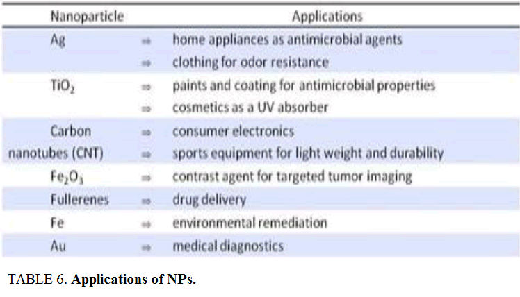
Discrete NMs are typically connected in nano-biomedicine as fluorescent natural labels and middle people for medication and additionally drug deliverance. Although this they can likewise be utilized for discovery of pathogens, tissue designing [7], tumor destruction, contrast change in (MRI) and phagokinetic examinations (Table 6).
Incomprehensible as antimicrobial
Amongst various metal NPs, AgNPs have been widely considered surgical gloves and covers, antibacterial injury dressings, bed lines and so forth [61]. They likewise have various applications in the fields of gadgets, catalysis and indicative therapeutics.
Biosensors
Similarly, gold nanoparticles (AuNPs) are additionally used for various purposes e.g. as labels for biosensors, for cure of hyperthermia and being equipped for conveying vast estimated biomolecules, give non-dangerous intends to quality and medication liberation to the objective locales [62].
Platinum nanoparticles (PtNPs) are utilized for the treatment of various illnesses, for example, growth and oxidative anxiety issue; in addition, they are utilized as a part of gadgets for planning of a novel memory component.
Environmental remediation
NMs offer the potential for the productive expulsion of toxins and natural contaminants from the earth. NMs capacity as adsorbents and catalysts in different shapes, for example, nanoparticles, tubes, wires, filaments and so forth and their composites with polymers are utilized for the discovery and evacuation of gasses, for example, (SO2, CO, NOx and so on.), violated chemicals (nitrate, arsenic, iron, substantial metals, manganese and so forth.), natural poisons (aliphatic and fragrant hydrocarbons) and characteristic substances, for example, (parasites, infections, microorganisms and anti-infection agents). Due to high surface region and high reactivity NMs demonstrate a superior execution in natural remediation than other regular procedures.
In sort to live in environment, containing high amounts of metals, life forms should have adjusted by advancing instruments to handle with them. These systems may include changing the potion way of the lethal metal so that it no more causes danger, bringing about the development of NPs of the metal concerned.
Clinical operations
These days in the clinical operations, in vivo sign of NPs in helpful capacities is a developing order. Although, there are still a few ambiguities with respect to their configuration and ramifications of NMs, for example, their pharmacokinetics, conveyance in the body, poisonous quality and security assessment earlier and consequent to their conjugation in therapeutic methods. Therefore, the biosimilarity of these NMs is still unclear and distinctive planning trials are in progress to maintain a considered distance from these injurious impacts. As yet, inferable from the nontoxic nature, AuNPs are broadly utilized as a part of healing and imaging operations.
Despite the fact that, it is impractical to make NMs that are non-hazardous to every single living cell, as there is no all-inclusive instrument for this reason, yet some essential examinations have been completed keeping in mind the end goal to make them more biocompatible and mitigate their cytotoxic impacts both in vitro and in vivo conditions.
Desired response
With the utilization of least conceivable dosage is typically regarded as an alluring answer for biocompatibility issues. Besides, the covering of NMs is additionally essential as broken, split or harsh packaging could trigger the safe reaction, in this way diminishing their bioavailability to the sought target locales [63].
Although worthy metals, NMs have developed as promising instruments against a standout amongst the most difficult human sickness i.e. tumor, however their definitive destiny in the body is still unclear with a specific end goal to interpret their general helpful potential.
cancer therapy

Figure 8: Applications of noble metal NPs for cancer therapy.
Optimization and Methodical Protocols
Optimization may topple reduce to the enhanced biosynthesis of NPs. Methodical protocols could be used for alloy of decidedly definite and well-characterized NPs right away on the quinine aspects, such as types of organisms, hereditary and genetical offering of organisms, incomparable publication for partition collecting and enzyme activity, optimal counteraction issuance and selection of the biocatalyst state have been considered. Morphology of the NPs depths is comforable by regulating the prior reaction conditions. Industrial scale combine of metal NPs gulp biomass needs diverse processes, in addition transmission enchant, insusceptibility of the transmit into the biomass, harvesting the cells make up of NPs by additionally metal ions to the cells, breaking of cells by classiness, homogenization of the cells to isolate the produced NPs, stabilization of the NPs, product formulation and quality control (Table 7).
Conclusion
Nanoparticles are having a great deal of utilizations in different fields like antimicrobials, additives, paints, biosensors and makeup. The use of microbes is the great way to deal with the creation of Eco-accommodating and costs adequate nanoparticles. Advancement of nanoparticles union by enhanced generation of society development will giving a decent prospect for the most extreme manufacture and it is exceptionally helpful for the numerous nanoparticles based applications. The utilization of microscopic organisms offers a method for creating "manufactories" for generation of nanoparticles essentially and reasonable. It is likewise apparent that NPs have extraordinary prospective in a wide range of businesses. The field of organic creation of nanoparticles is moderately new and underexplored; in any case it demonstrates awesome potential in the biotechnology part.
Future Prospects
The research work clearly predicted the NPs synthesis abilities of bacteria; nonetheless, it could be enhanced as far as rate and quality by exploring proficient microorganisms which are safe and expressive against metals by delivering certain synergist proteins activated through various qualities.
Assessment and comprehension the fundamental systems in term of biomolecules required in changing and balancing out the metal particles into Nano-composites.
Choose the best bacteria, which have the best growth rate and characteristics to synthesize NPs with specific and smaller shapes.
Scaling up the laboratory methods to the industrial scale that may lead to the enhanced synthesis of NPs.
References
- Sintubin L, Windt W, Dick J, et al. Lactic acid bacteria as reducing and capping agent for the fast and efficient production of silver nanoparticles. ApplMicrobiolBiotechnol. 2009;84:741-9.
- Jadhav JP, Kalyani DC, Telke AA, et al. Evaluation of the efficacy of a bacterial consortium for the removal of color, reduction of heavy metals and toxicity from textile dye effluent. Biores Technol. 2010;101:165-73.
- Bazylizinki DA, Heywood BR, Mann S. Fe304 and Fe3S4 in a bacterium. Nature. 1993;366:218.
- Sweeney RY, Mao C, Gao X, et al. Bacterial biosynthesis of cadmium sulfide nanocrystals.ChemBiol. 2004;11(11):1553-9.
- Bai H, Zhang Z, Gong J. Biological synthesis of semiconductor zinc sulfide nanoparticles by immobilized Rhodobactersphaeroides. BiotechnolLett. 2006;28(14):1135-39.
- Wei X, Luo M, Li W, et al. Synthesis of silver nanoparticles by solar irradiation of cell-free Bacillus amyloliquefaciens extracts and AgNO3. Bioresour Technol. 2012;103:273-8
- Baesman SM, Bullen TD, Dewald J, et al.Formation of tellurium nanocrystals during anaerobic growth of bacteria that use Te oxyanions as respiratory electron acceptors. Appl Environ Microbiol. 2007;73(7):2135-43.
- Bharde A, Wani A, Shouche Y, et al. Bacterial aerobic synthesis of nanocrystalline magnetite. J Am Chem Soc. 2005;127(26):9326-7.
- Mukherjee P, Ahmad A, Mandal D, et al. Bioreduction of AuCl(4)(-) ions by the fungus, verticillium sp. and surface trapping of the gold nanoparticles formed D.M. and S.S. thank the Council of Scientific and Industrial Research (CSIR), Government of India, for financial assistance. AngewChemInt Ed Engl. 2001;40:3585-8.
- Bansal V, Poddar P, Ahmad A. et al. Room-temperature biosynthesis of ferroelectric barium titanate nanoparticles. J Am Chem Soc. 2006;128:11958-63.
- Cai J, Kimura S, Wada M, et al. Nanoporous cellulose as metal nanoparticles support. Biomacromolecules. 2009;10:87-94.
- Hofmann H. Nanomaterials; Introduction and Atoms Cluster. 2009.Version 2.0 09.2011.
- Wei X, Luo M, Li W, et al. Synthesis of silver nanoparticles by solar irradiation of cell-free Bacillus amyloliquefaciensextracts and AgNO3. Bioresour Technol. 2012; 103:273-8
- Kalimuthu K, Suresh BR, Venkataraman D, et al.Biosynthesis of silver nanocrystals by Bacillus licheniformis. Colloids Surf B Biointerfaces. 2008;65:150-3.
- Konishi Y, sukiyama T, Ohno K, et al. Intracellular recovery of gold by microbial reduction of AuCl-4ions using the anaerobic bacterium Shewanella algae.Hydrometallurgy. 2006;81(1):24-9.
- Mokhtari N, Daneshpajouh S, Seyedbagheri S, et al. Biological synthesis of very small silver nanoparticles by culture supernatant of Klebsiella pneumonia: The effects of visible-light irradiation and the liquid mixing process. Mater Res Bull; 2009;44(6):1415-21.
- Rai M, Yadav A, Gade A. Silver nanoparticles as a new generation of antimicrobials. Biotech Adv. 2009;27:76-83.
- Sunkar S, Nachiyar CV. Biogenesis of antibacterial silver nanoparticles using the endophytic bacterium Bacillus cereus isolated from Garciniaxanthochymus. Asian Pac J Trop Biomed. 2012;2(12)953-9.
- Sanpo N, Wen C, Berndt CC, et al. Antibacterial properties of spinelferrite nanoparticles. In: Mendez A, editor. Microbial pathogens andstrategies for combating them: Science, technology and education.Spain: Formatex Research Centre. 2013;239-50.
- Beveridge TJ, Murray RGE. Sites of metal deposition in the cell wall of Bacillus subtilis. J Bacteriol. 1980;141(2):876-87.
- Mahanty A, Bosu R, Panda P, et al. Microwave assisted rapid combinatorial synthesis of silver nanoparticles using E. coli culture supernatant.Inter J Pharma Bio Sci 2013;4(2):1030-5.
- Shahverdi AR, Minaeian S, Jamalifar H, et al. Rapid synthesis of silver nanoparticles using culture supernatants of Enterobacteria: A novel biological approach. Process Biochemistry. 2007;42:919-23.
- Manivasagan P, Venkatesan J, Senthilkumar K, et al. Biosynthesis, antimicrobial and cytotoxic effect of silver nanoparticles using a novel Nocardiopsissp. MBRC-1. BioMed Res Inter. 2013;287638-9
- Lengke M, Southam G. Bioaccumulation of gold by sulfate-reducing bacteria cultured in the presence of gold (I)- thiosulfate complex. Geochimica et CosmochimicaActa; 2006;70(14):3646-61.
- Lengke, MF, Southam G. The effect of thiosulfate oxidizing bacteria on the stability of the gold-thiosulfate complex. Geochimica et CosmochimicaActa. 2005;69(15):3759-72.
- Bazylinski DA, Frankel RB, Jannasch HW. Anaerobic magnetite production by a marine, magnetotactic bacterium. Nature. 1988;334:518-9.
- Holmes JD, Smith PR, Gowing RE, et al.Energy-dispersive X-ray analysis of the extracellular cadmium sulfide crystallites of Klebsiella aerogenes. ArchMicrobiol. 1995;163(2):143-7.
- Gerrard TL, Telford JN, Williams HH. Detection of selenium deposits in Escherichia coli by electron microscopy. J Bacteriol. 1974;119(3):1057-60.
- Kumar V, Yadav SK. Plant-mediated synthesis of silver and gold nanoparticles and their applications. J ChemTechnolBiotechnol. 2009;84(2):151-7.
- Das V, Thomas R, Varghese R, et al. Extracellular synthesis of silver nanoparticles by the Bacillus strain CS 11 isolated from industrialized area. 3 Biotech. 2014;4:121-6.
- Kalishwaralal K, Deepak V, Ramkumarpandian S, et al. Extracellular biosynthesis of silver nanoparticles by the culture supernatant of Bacillus licheniformis. Mat Lett. 2008;62(29):4411-3
- Woolfolk CA, Whiteley HR. Reduction of inorganic compounds with molecular hydrogen by Micrococcus lactilyticus. I. Stoichiometry with compounds of arsenic, selenium, tellurium, transition and other elements. J Bacteriol. 1962; (84):647-58.
- Bhowmik D, Chiranjib, Chandira RM, et al. Nanomedicine-an overview. Int JPharm Tech Res. 2010;2(4):2143-51.
- Bai HJ, Zhang ZM, GuoY, et al. Biosynthesis of cadmium sulfide nanoparticles by photosynthetic bacteria Rhodopseudomonas palustris. Coll Surf BBiointerf. 2009;70(1):142-146.
- Oremland RS, Herbel MJ, Blum JS, et al. Structural and spectral features ofselenium nanospheres produced by Se-respiring bacteria. Appl Environ Microbiol. 2004;70(1):52-60.
- Kasthuri J, Kathiravan K, Rajendiran N. Phyllanthin-assisted biosynthesis of silver and gold nanoparticles: A novel biological approach. J Nanopart Res. 2008;11:1075-85.
- Park Y, Hong YN, Weyers A, et al. Polysaccharides and phytochemicals: A natural reservoir for the green synthesis of gold and silver nanoparticles. IET Nanobiotechnol. 2011;5:69-78.
- Narayanan KB, Sakthivel N. Biological synthesis of metal nanoparticles by microbes. Adv Colloid Interface Sci. 2010;156:1-13.
- Raveendran P, Fu J, Wallen SL. Completely “green” synthesis and stabilization of metal nanoparticles. J Am Chem Soc. 2003;125:13940-1.
- Beveridge JT, Hughes MN, Lee H, et al. Metal-microbe interactions: Contemporary approaches. Advances in MicrobialPhysiology. 1997;38:178-243.
- Mohanpuria P, Rana NK, Yadav SK, et al. Biosynthesis of nanoparticles: Technological concepts and future applications.J Nanopart. 2008;10(3):507-17.
- Angell P. Understanding microbially influenced corrosion as biofilm-mediated changes in surface chemistry. CurrOpin Biotech. 1999;10(3):269-72.
- Zierenberg RA, Schiffman P. Microbial control of silver mineralization at a sea-floor hydrothermal site on the northern Gorda Ridge. 1990;348(6297):155-7.
- Harvey PI, Crundwell FK. Growth of Thiobacillusferrooxidans: A novel experimental design for batch growthand bacterial leaching studies. Appl Environ Microb. 1997;63(7):2586-92.
- Stephen JR, Macnaughton SJ. Developments in bacterial remediation of metals.CurrOpin Biotech. 1990;10(3):230-3.
- Kang SH, Bozhilov KN, Myung NV, et al. Microbial synthesis of CdS nanocrystals in genetically engineered E.coli. AngewChemInt Edit, 2008;47(28):5186-9.
- Deplanche k, Macaskie V, Biorecovery of gold by Escherichia coli and Desulfovibriodesulfuricans. Biotech Bioeng. 2008;99(5):1055-64.
- Michel C, Brugna M, Aubert C, et al. Enzymatic reduction of chromate: comparative studies using sulfate-reducing bacteria: Key role of polyheme cytochromes C and hydrogenases. ApplMicrobiolBiot. 2001;55(1):95-100.
- Zadvorny OA, Zorin,Gogotov IN. Transformation of metals and metal ions by hydrogenases from phototrophic bacteria.Arch MicrobioL. 2006;184(5):279-85.
- Matsunaga T, Takeyama H. Biomagnetic nanoparticle formation and application.Supramol Sci. 1998;5(4):391-4.
- Arakaki A, Nakazawa H, Nemoto M, et al. Formation of magnetite by bacteria and its application. J R Soc Interface. 2008;5(26):977-99.
- Moisescu C, Bonneville S, Tobler D, et al. Controlled biomineralization of magnetite (Fe3O4) byMagnetospirillumgryphiswaldense.MineraL Mag. 2008; 72(1):333-6.
- Satomi M,Oikawa H, Yano Y.Shewanellamarinintestina sp. nov., Shewanellaschlegeliana sp. nov.andShewanellasairae sp. nov., novel eicosapentaenic-acid-producing marine bacteriaisolated from sea-animal intestines.Int J SystEvolMicr 2003;53(2):491-9.
- Moreau JW, Weber PK, Martin MC, et al. Extracellular proteins limit the dispersal of biogenic nanoparticles. 2007;316(5831):1600-3.
- Amemiya Y, Arakaki A,Staniland SS, et al. Controlled formation of magnetite crystal by partial oxidation of ferrous hydroxide in the presence of recombinant magnetotactic bacterial protein Mms6.Biomaterials. 2007;28(35):5381-9.
- Prozorov T, Mallapragada SK, Narasimhan B, et al. Proteinmediated synthesis of uniform superparamagnetic magnetite nanocrystals. AdvFunct Mater. 2007;17(6):951-7.
- Prozorov T, Palo P, Wang L, et al. Cobalt ferrite nanocrystals: Out-performing magnetotactic bacteria. ACS Nano. 2007;1(3):228-33.
- Bankinter Foundation. Nanotechnology; The industrial revolution of the 21st century. 2006.
- Lee H, Purdon AM, Chu V, et al. Controlled assembly of magnetic nanoparticles from magnetotactic bacteria using microelectromagnets arrays. Nano Lett. 2004;4:995-8.
- Panigrahi S, Kundu S, Ghosh SK, et al. Selective one-pot synthesis of copper nanorods under surfactantless condition. Polyhydron. 2006;25(5):1263-9.
- Bellucci S. Nanoparticles and nanodevices in biological applications. Lecture Notes in Nanoscale Sci Technol. 2009;4:1-198.
- Acosta TLS, Lopez MLM, Nunez ARE, et al. Biocompatible metal-oxide nanoparticles:Nanotechnology improvement of conventional prosthetic acrylic resins. J Nanomater. 2011:941561.
- Driscoll TJ, Lawandy NM, Nouri M, et al. Conferenceon Lasers and Electro-Optics; Baltimore, MD, USA:Optical Society of America. 1997.
- Salata O. Applications of nanoparticles in biology and medicine. J Nanobiot. 2004;2(1):3.
- Contado C, Pagnoni A. TiO2 in commercial sunscreen lotion: Flowfield-flow fractionation and ICP-AES together for size analysis. Anal Chem. 2008;80(19):7594-608.
- Guo Y, Zhou Y, Jia D, et al. Fabrication and in vitrocharacterizationof magnetic hydroxycarbonate apatite coatings with hierarchicallyporous structures. ActaBiomater. 2008;4(4):923-931.
- Sikong L, Kooptarnond K, Niyomwas S, et al. Photoactivityand hydrophilic property of SiO2 and SnO2 co-doped TiO2 nanocompositethin films. Songklanakarin Journal of Science & Technology. 2010;32(4):413-8.
- Arbab AS, Bashaw LA, Miller BR, et al. Characterization of biophysicaland metabolic properties of cells labeled with superparamagneticironoxide nanoparticles and transfection agent for cellular MR imaging. Radiology. 2003;229(3):838-46.
- Limbach LK, Wick P, Manser P, et al. Exposure of engineered nanoparticles to human lung epithelial cells:Influence of chemical composition and catalytic activity on oxidativestress. Environ Sci Technol. 2007;41(11):4158-63.
- Nino MN, Martínez CGA, Aragon PA. Characterizationof silver nanoparticles synthesized on titanium dioxide fine particles. Nanotech. 2003;19(6):065711.
- Ahn SJ, Lee SJ, Kook JK. Experimental antimicrobial orthodonticadhesives using nanofillers and silver nanoparticles. Dent Mater. 2009;25(2):206-13.