Original Article
, Volume: 15( 2)Characterization and Antimicrobial Activity of Green Synthesized Zinc Sulphide Nanoparticles Using Plant Extracts of Phyllanthus niruri
- *Correspondence:
- Sathish Kumar M Thin Film R&D centre, Department of Electronics, Erode Arts and Science College, Erode, Tamil Nadu, India
Tel: +91-9865861230; E-mail: mskeasc@gmail.com
Received Date: May 08, 2017 Accepted Date: May 12, 2017 Published Date: May 16, 2017
Citation: Sathish Kumar M, Saroja M, Venkatachalam M. Characterization and Antimicrobial activity of Green Synthesized Zinc Sulphide Nanoparticles using Plant Extracts of Phyllanthus niruri. Int J Chem Sci 2017;15(2):134.
Abstract
We report a biosynthesis zinc sulphide nanoparticles using plant extract of Phyllanthus niruri and their antimicrobial activity. Biosynthesized ZnS Nanoparticles were excellent crystalline structure formation confirmed by using X-ray powder diffraction, scanning electron microscopy use to identify surface morphology of ZnS Nanoparticles, FT-IR used to found different functional group of biosynthesized ZnS NP’s and Optical absorbance were evaluate by UV-Visible Spectrometer. The antimicrobial activity of biosynthesis ZnS nanoparticles using Phyllanthus niruri methanol plant extract were determined by disc diffusion method against various pathogenic microorganisms.
Keywords
ZnS; Antimicrobial Activity; Phyllanthus niruri; XRD; FTIR
Introduction
Synthesis of Nanoparticles were significant field in nanotechnology because of their size, shape, dimension and the different properties like a magnetic, mechanical, thermal, electronic and high surface area are used to develop nanotechnology [1,2]. In the field of nanotechnology diverse form of nanoparticle like a metal [3] carbon [4-7] polymers [8] are available, chemical methods and chemical co-operation [9], sol-gel [10] methods are mostly used to synthesis nanoparticles. In the field of nanotechnology various applications used here antimicrobial activity is one of the application [11,12] widely gold, silver nanoparticles used in food and healthcare industries. Zinc sulphide is II-IV group semiconductor (3.68 eV) is a wide band gap energy materials so its suiTable for biosensors, fluorescent, chemical sensors [13], Doped ZnS nanoparticles are good antibacterial agents [14], Other sulphide and oxide materials widely investigated and compare for antimicrobial applications [15,16]. ZnS is an inorganic material so used useful practical applications [17,18], This study was first time report biosynthesizing zinc sulphide nanoparticles using Pyllanthusniruri plant extract and their antimicrobial activity against various pathogenic microorganisms.
Materials and Methods
Plant material extraction
Well matured fresh Phyllanthus niruri plant were collected around idappadi waste land and the Phyllanthus niruri plant materials washed three times in tap water and two times in DI water at room temperature during the process all dust and soils are removed and collected plants are dried at room temperature in open air, shadow dried ten days. Then 50 g of coarse powder was crushed dried plant material by using grinder, the soxhlet apparatus were used to plant extraction of 250 ml methanol solvent using coarse powder and extracts was used to treat further investigation.
Method of preparation ZnS nanoparticles
Zinc sulphate (14.4 g) were dissolved in 100 ml DI water and treated 30 minutes contentious stirring at 400°C, 1 mm zinc sulphate aqueous solution was prepared. Then 20 ml of Phyllanthus niruri methanol plant extracts was added with 1 mm (80 ml) prepared zinc sulphate solution drop by drop and mixing treated room temperature at 2 hours while continuous stirring and result were formed zinc sulphide nanocolloid solution that’s treated 600°C at 5 hours for drying.
Inoculums preparation for antibacterial activity
Muller Hinton broth (MHB) test plate was incubated for 24 hours at 25°C to 370°C and loopful cell transferring process is good for active culture, 40°C is good temperature for growth stock culture slope nutrient agar. The optical densities of bacterial 2.0 × 10⁶ colony forming units (CFU/ml) were diluted in MHB plates.
Antimicrobial susceptibility test
Muller Hinton Agar (MHA) plates were used to analysis antimicrobial activity of invitro disc diffusion method and plate obtained from hi-media Mumbai. Te normal MHA plates allow to pouring process using 15 ml molten media and allow 5 minutes for solidify then 0.1% inoculums culture was coated uniformly surface of the plate and dry 5 min, the different concentration 60 mg disc plates was placed in surface of MHA plates width of the disc is 6 mm. then the synthesis plant extract was loaded at different concentration on the disc and allow 5 min extracts was diffuse the disc, loaded MHA plates were kept 37°C for 24 hours in incubation. During the incubation, the maximum zone of inhibition was formed around the disc and transparent millimeter ruler used to measure inhibition zone.
Phytochemical screening test
Mayer’s reagent and Wagnr’s reagent used to analysis of alkaloids, the Phyllanthus niruri plant extract was added and filtered with HCL acid. At mayer regents test yellow color formation was indicate an alkaloid, brown color formation conform alkaloid in wagner’s reagent. H2S04were added with lead acetate and plant extract is added drop by drop, then the yellow color formation was found and its clearly indicate the presence of flavonoids. 1 ml of acetic anhydride added with H2S04 and plant extract which changes the color violet in to blue or green it’s conform presence of steroids. The terpenoids presence is confirmed by Salkowski test when chloroform mixed together with H2S04 and plant extract the reddish-brown color was formed. Anthrauinones confirmed at borntrager’s test, extract boiled in HCL and cooled with water bath then ammonia solution added drop by drop with CHCl3 the result was formed pink color formation. The block and yellow color formation was conformed phenol were ferric chloride and lead acetate mixed drop by drop in plant extract solution. The plant extract reacts with DI water during shaken the dark block color was formed from green color it confirms saponins presence. Tannins presence conformed by adding ferric chloride with heat water and plant extract it was change dark green color in to black color. Carbohydrate, oil and resins was found by using DI water and plant extract, the extract and DI waters are shaken and filtered by using filter paper.
Characterization techniques
The synthesized ZnS crystalline structure was calculated by X-ray diffractometer with CuKa1 Radiation (α=1.5405Å) in (XPERT-PRO). Hitachi S-4500 (SEM) is used to find surface roughness. JASCO FT/1 IR-6600 instrument was carried out the Fourier transform infrared spectroscopy (FT-IR) measurement and optical absorbance spectra were taken using UV-Visible Spectrometer.
Results and Discussion
X-ray diffraction pattern
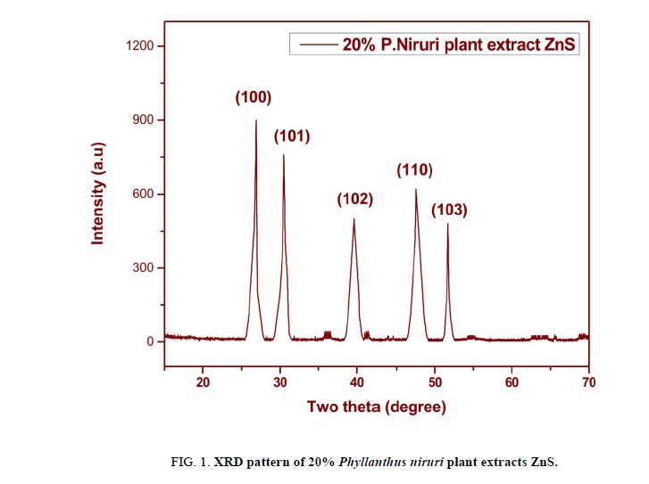
Biosynthesized ZnS Nanoparticles crystalline power sample structure was studied by using XRD, the unique peak (2θ) of 26.91, 30.52, 39.61, 47.56, 51.77 degrees, corresponding reflection plane is 100,101,102,110 and 103 respectively its shown Figure. 1. The strong intensity peak at 26.91 degree in (100) plane its conformed ZnS Hexagonal wurtzite synthesis structure of ZnS (JCPDS: 36-1450).
Scherrer’s formula used to calculate average grain size of Phyllanthus niruri plant extract ZnS nanoparticles.
D=0.9 λ / β cos ? -------------------(1)
Where λ=1.5405 Å for CuKa1, ? is Bragg’s angle, β is (FWHM) full of half maximum of corrected instrumental peak in radian, the average crystalline size of the zinc sulfide was found to be ~74 nm respectively.
Morphology studies
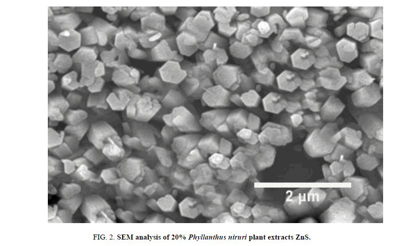
The surface morphology of Phyllanthus niruri plant extracts ZnS nanoparticles observed by scanning electron microscopy (SEM), it is a versatile technique for studying microstructure. Prepared biosynthesized ZnS NP’s power sample examined and found different hexagonal shape and size to be less than 2 μm it’s clearly shows Figure. 2.
FT-IR Spectrum analysis
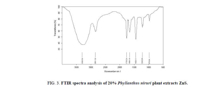
Phyllanthus niruri Plant extracts ZnS nanoparticles has different function group carried out to identify Furrier Transferred infrared (FTIR) was shown in Figure. 3 range from 4000-400 cm-1 wavenumbers. The peak at 3300 cm-1 is Secondary amines (O-H) band of the Phyllanthus niruri, peak transfer to the 2851 cm-1 band it indicates alkanes (C-H) bond, another (C=O) Zinc bond at 1739 cm-1 in very strong stretching, 1685 cm-1 at aromatic ketones (C==C) Zinc sulphide bond very strong stretching.
At the edge of 1426 cm-1 Aliphatic aldehydes (C=H) a strong peak at indicate Zinc bond, Tertiary butyl 1240 cm-1 (O=H) strong stretching, cycloalkanes (C=C) medium stretching at 978 cm-1 indicate the formation of nanoparticles.
UV-visible analysis
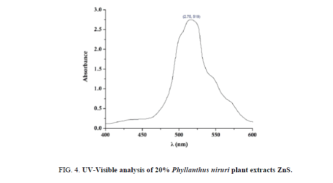
UV-Visible analysis of Phyllanthus niruri plant extract ZnS nanoparticles clearly shown in Figure. 4, The optical absorbance range from 400-600 nm, Then the maximum absorbance of Phyllanthus niruri plant extract ZnS nanoparticles colloid solution was fall in at 519 nm It is found that the absorption edge shifts toward longer wavelength in biosynthesized ZnS.
Antimicrobial activity of ZnS
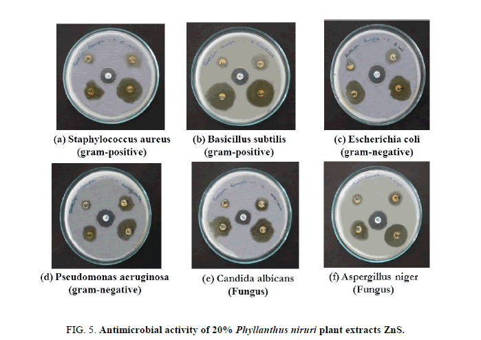
Qualitative phytochemical analysis of Phyllanthus niruri plant extract ZnS was found Alkaloids, Flavonoids, Steroids, Phenols, Tanin, Carbohydrates were absence in Terpenoids, Arthroquinone, Saponin, and Oil and Resins. In this phytochemical analysis found Phyllanhus niruri plant extract mixed with ZnS colloidal solution and this was treated in the different concentration (Table 1).
| Organism | C | 30 µl | 40 µl | 50 µl | 60 µl |
|---|---|---|---|---|---|
| S. aureus | 17 | 11 | 16 | 19 | 26 |
| B. subtilis | 16 | 10 | 14 | 19 | 22 |
| E. coli | 19 | 14 | 18 | 23 | 29 |
| P. aerugoinosa | 21 | 10 | 14 | 18 | 28 |
| C. albicans | 18 | 12 | 24 | 26 | 32 |
| A. niger | 20 | 14 | 16 | 18 | 22 |
Table 1. Antimicrobial activity of Phyllanthus niruri plant extracts ZnS.
Muller Hinton Agar (MHA) plates were used to analysis antimicrobial activity of in vitro disc diffusion method and plate obtained from hi-media Mumbai. Te normal MHA plates allow to pouring process using 15 ml molten media and allow 5 minutes for solidify then 0.1% inoculums culture was coated uniformly surface of the plate and dry 5 min, the different concentration 60 mg disc plates was placed in surface of MHA plates width of the disc is 6 mm. then the synthesis plant extract was loaded at different concentration on the disc and allow 5 min extracts was diffuse the disc, loaded MHA plates were kept 37°C for 24 hours in incubation. During the incubation, the maximum zone of inhibition was formed around the disc and transparent millimeter ruler used to measure inhibition zone that clearly shown in Figure. 5 and Table 1 Here bacteria and fungus are tested S. aureus, B. subtilis are gram positive E. coli and P. aeruginosa gram negative bacteria, C. albicans, A. niger as fungus culture. At concentration 30 μl the high antimicrobial zone was formed around (14 mm) in E. coli and N. niger followed by C. albicans (12 mm), in S. aureus (11 mm), B. subtilis and P. aeruginosa affected only (10 mm) its low zone compare other organisms. In 40 μl and 50 μl concentration C. albicans followed by E. coli has high zone of inhibition around 24 mm and 26 mm. Finally at concentration 60 μl good inhibition zone was formed in all tested microorganisms, maximum antimicrobial zone were formed in C. albicans (32 mm) of fungus culture followed by gram negative and positive bacteria’s E. coli (29 mm), P. aeruginosa (28 mm), S. aureus (26 mm) and small inhibition zone found in B. subtilis and N. niger (22 mm) so the biosynthesized ZnS NPs using Phyllanthus niruri plant extract was excellent antimicrobial activity against all tested microorganisms like gram positive, gram negative and fungus cultures.
Conclusion
Here we reported green synthesis of ZnS NPs using Phyllanthus niruri plant extract first time, the characterization of ZnS NPs was analysis by using XRD, SEM, FTIR, UV-Visible. ZnS NPs are Hexagonal Wurtzite structure and average crystalline size was calculated ~74 nm using XRD powder diffraction, SEM used to analysis different hexagonal size of the nanoparticles surface it found below 2 μm. To identifying different functional group of Phyllanthus niruri plant extract synthesized ZnS NPs were using FTIR. UV-Visible maximum optical absorbance frequency was absorbed in 519 nm. The in vitro disc diffusion method used to analysis antimicrobial activity of ZnS NPs and the result shows excellent inhibition zone was formed against all tested organisms. Here maximum inhibition zone was observed in Candida albicans (32 mm) fungus culture, Minimum inhibition zone was found in Basicillus subtilis (22 mm) gram positive bacteria followed by Aspergillus niger (22 mm) fungus at concentration of 60 μl. So, it was the evident the biosynthesized ZnS NPs using Phyllanthus niruri plant extracts have excellent Antimicrobial activity and its used different bioelectronics applications.
References
- Manojkumar K, Sivaramakrishna A, Vijayakrishna K. A short review on sTable metal nanoparticles using ionic liquids, supported ionic liquids and poly (ionic liquids). J Nanopart Res. 2016;18:1-22.
- Chang CC, Chiang FC, Chen SM, et al. Studies on electrochemical oxidation of aluminum and dyeing in various additives towards industrial applications. Int J Electro chem Sci. 2016;11:2142-52.
- Palanisamy S, Thangavelu K, Chen SM, et al. Preparation and characterization of gold nanoparticles decorated on grapheneoxide polydopamine composite: Application for sensitive and low potential detection of catechol. Sens Actuators. B Chemical. 2016;233:298-306.
- Palanisamy S, Thangavelu K, Chen SM, et al. Preparation and characterization of a novel hybrid hydrogel composite of chitin stabilized graphite: Application for selective and simultaneous electrochemical detection of dihydroxybenzene isomers in water. J Elec anal Chem. 2017;785:40-7.
- Kokulnathan T, Palanisamy S, Chen SM, et al. Electrochemical determination of caffeic acid in wine samples using reduced graphene oxide/polydopamine composites. J Electro chem Soc. 2016;163:B726-B31.
- Palanisamy S, Kokulnathan T, Chen SM, et al. Preparation of chitosan grafted graphite composite for sensitive detection of dopamine in biological samples. Carbohydrate Polymers. 2016;151:401-7.
- Thirumalraj B, Palanisamy S, Chen SM, et al. A simple electrochemical platform for detection of nitrobenzene in water samples using an alumina polished glassy carbon electrode. J coll inter sci. 2016;475:154-60.
- Palanisamy S, Kokulnathan T, Chen SM, et al. Synthesis and characterization of polypyrrole decorated graphene/β-cyclodextrin composite for low level electrochemical detection of mercury (II) in water. Sensors and Actuators B: Chemical. 2017;243:888-94.
- Ameer A, Faheem A, Nishat A, et al. Biosynthesis and characterization of ZnO nanoparticles using (Lactobacillus plantarum). J Alloys Comp. 2010;496:399-402.
- Alessio B. Maximilian D, Pierandrea LN, et al. Synthesis and characterization of zinc oxidenanoparticles: application to textiles as UV-absorbers. J Nanopart Res. 2011;10:679-89.
- Pelgrift RY, Friedman AJ. Nanotechnology as a therapeutic tool to combat microbial resistance. Adv Drug Deliv Rev. 2013;65:1803-15.
- Bokare A, Sanap A, Pai M, et al. Antibacterial activities of Nd doped and Ag coated TiO2 nanoparticles under solar light irradiation. Colloids Surf B. 2013;102:273-80.
- Fang X, Zhai T, Gautam UK, et al. ZnS nanostructures from synthesis to applications. Prog Mater Sci. 2011;56:175-287.
- Singh P, Sharma PK, Kumar M, et al. Redluminescent manganese-doped zinc sulphide nanocrystals and their antibacterial study. J Mater Chem B. 2014;2:522-8.
- Malarkodi C, Annadurai G. A novel biological approach on extracellular synthesis and characterization of semiconductor zinc sulfide nanoparticles. Applied Nanoscience. 2013;3:389-95.
- Vanaja M, Rajeshkumar S, Paulkumar K, et al. Phytosynthesis and characterization of silver nanoparticles using stem extract of Coleus aromaticus. Int J Mat Biomat App. 2013;3:1-4.
- Malarkodi C, Chitra K, Rajeshkumar S. Novel ecofriendly synthesis of titaniumoxide nanoparticles by using Plano micro biums pand its antimicrobial evaluation. Der Pharmacia Sinica. 2013;4:59-66.
- John R, Sasiflorence S. Optical, structural and morphological studies of bean like ZnS nanostructures by aqueous chemical method. Chalcogenide Let. 2010;27:269.
